The objective of the 3D analysis provided by Visible Patient is to give, from a medical image, a fast, quantitative and precise measurement of the volume of damaged lung.
From a simple X-ray of the patient, Visible Patient extracts a 3D digital clone of the lungs. The advantage of this analysis is linked to the precise delineation of each infected or healthy region of the lung. It allows to define the remaining functional volume as well as the damaged volume, in particular an area referred to as “ground glass” which is systematically found in case of Covid-19 infection. From the value of those volumes our project has defined a new diagnosis of severity that has to be validated.
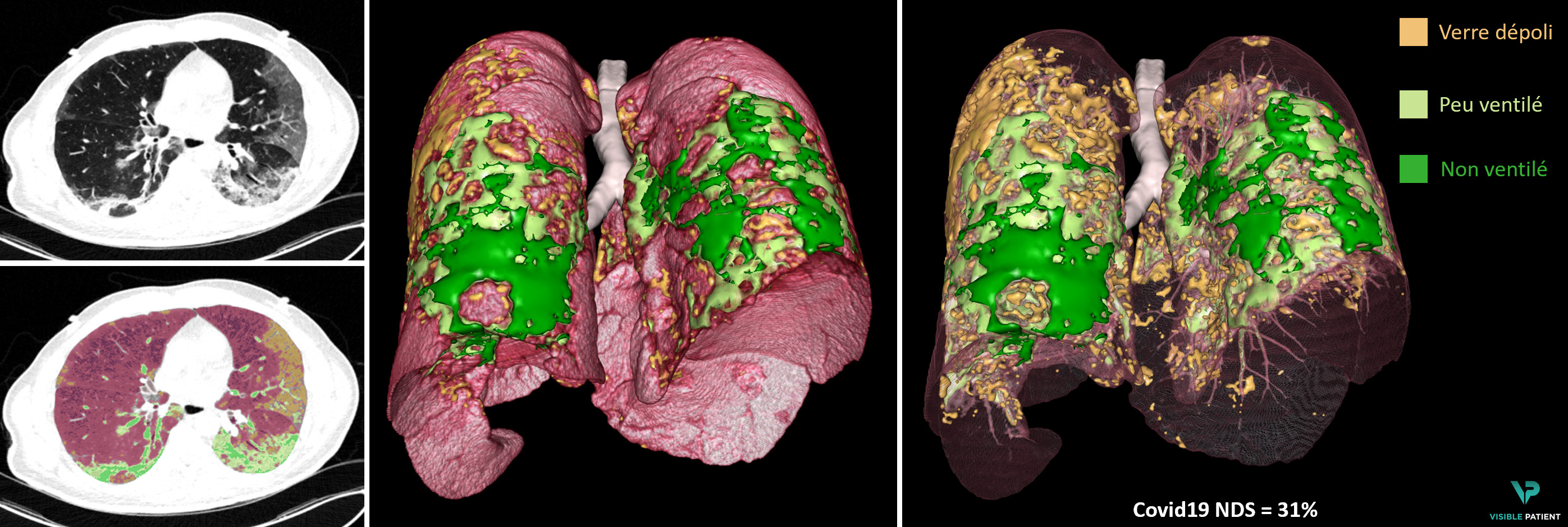 From an X-ray scan (left), result of the 3D analysis (in posterior view) by Visible Patient providing the 3D model of pathological areas (orange and green) and the new diagnosis of severity NDS, here 31%.
For Research/Investigational Use only. The safety, effectiveness and performance characteristics of the New Diagnosis of Severity score related to the severity of lung damage induced by COVID-19 described herein, as well as the results, have not been established and is under scientific evaluation.